Collagen injection to the breasts

Collagen injection to the breasts
18Nov29
A female presented with a dominant mass on her left breast. She has history of collagen injection on her breast before. I requested for an ultrasound hoping it could help in the diagnosis of the dominant mass. However, it didn’t. Report just said obscured breasts (by collagen). I decided to do needle evaluation and aspiration. On aspiration, I was able to aspirate with difficulty 9 cc of oily substance, which I assumed to be the collagen injected before and the dominant mass decreased in size.
Personal advice: Do not go for injectables for breast augmentation like collagen and silicone. You will regret it in the future because it will create problems such as granuloma (chronic inflammation causing dominant mass) and difficulty to screen for breast cancer.

PLS. DON’T. I CARE FOR YOU.

IMG_1287


IMG_1295

IMG_1289

Posted in Breast Augmentation | Leave a comment

Locally Advanced Breast Cancer – Rhomboid Flap Reconstruction

Locally Advanced Breast Cancer – December 2014

No response (20%) to neoadjuvant chemotherapy – 3 cycles of CMF

Total mastectomy with axillary dissection with rhomboid flap reconstruction – April 6, 2015

December 2014

amy_calatrava_14dec27 (1)

February 2015

amy_calatrava_15fe21 (1)

March 2015

amy_calatrava_15mar13 (1)

April 2015

breast_ca_amy_calatrava_15apr6 (1)

breast_ca_amy_calatrava_15apr6 (2)

breast_ca_amy_calatrava_15apr6 (4)

breast_ca_amy_calatrava_15apr6 (7)

Posted in Locally Advanced Breast Cancers | Leave a comment

2013 in review

The WordPress.com stats helper monkeys prepared a 2013 annual report for this blog.

Here’s an excerpt:

A San Francisco cable car holds 60 people. This blog was viewed about 980 times in 2013. If it were a cable car, it would take about 16 trips to carry that many people.

Click here to see the complete report.

Posted in Review of ROJoson's Blogs in Wordpress.com | Leave a comment

Preventing Surgical Complications of Modified Radical Mastectomy – 2013

ROJoson’s Lecture in 2013 Postgraduate Course – Department of Surgery, Philippine General Hospital

Preventing Surgical Complications of Modified Radical Mastectomy – Improving Outcomes – How I Usually Do it

http://www.slideshare.net/rjoson/preventing-complications


Posted in Mastectomy Morbidities | Leave a comment

Mastectomy Morbidities: Prevention, Detection, and Treatment (Focus: Seroma, Infection, Bleeding)

Mastectomy Morbidities: Prevention, Detection, and Treatment (Focus: Seroma, Infection, Bleeding)

ROJoson’s lecture in the 2008 UP-PGH Department of Surgery Postgraduate Course.

Posted in Mastectomy Morbidities | Leave a comment

Keloids and Mastectomy Incision Planning

Keloids and Mastectomy Incision Planning

The situation of the keloids in the sternal area that may influence the direction of the elliptical mastectomy incision:

“Keloids can crop up anywhere but do so most easily on certain areas, such as the skin around the upper chest and shoulders – particularly over the breastbone (sternum) – and on the earlobes.”

 http://www.bad.org.uk/site/834/default.aspx

Sites for keloid formation: the most common areas are the sternum, shoulder, earlobe and cheek.

http://www.patient.co.uk/doctor/Keloid-Scars.htm

·  Al-Attar A, Mess S, Thomassen JM, et al; Keloid pathogenesis and treatment. Plast Reconstr Surg. 2006 Jan;117(1):286-300.

·  Leventhal D, Furr M, Reiter D; Treatment of keloids and hypertrophic scars: a meta-analysis and review of the literature. Arch Facial Plast Surg. 2006 Nov-Dec;8(6):362-8.

Certain areas of the body such as the sternum, deltoid region of the upper arm, and upper back, have increased susceptibility to keloid formation.

http://emedicine.medscape.com/article/876214-overview#aw2aab6b3

Based on the above situations, the risk for keloid in mastectomy scars is at least theoretically higher in those incisions that approach the sternal area, particularly the upper part.  Thus, a recommendation will be to avoid this area and place the medial part of the mastectomy incision on the lower part of the sternum.   Thus, an oblique elliptical mastectomy incision should be preferred over a transverse elliptical mastectomy incision if the risk of keloid formation is to be avoided.   (Note: Above statements have to be validated with a clinical research.)

Note: Other factors have to be considered before deciding on the direction of the mastectomy incision.

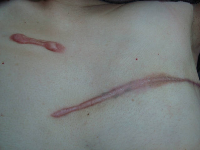
Pictures of patients with keloid on the sternum.DSC04604

DSC04348

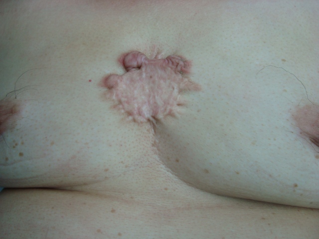
Keloids before modified radical mastectomy for breast cancer on the left.

DSC05152

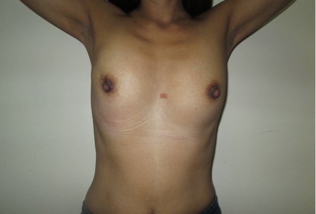
A month after the mastectomy using an oblique mastectomy incision.

DSC04504

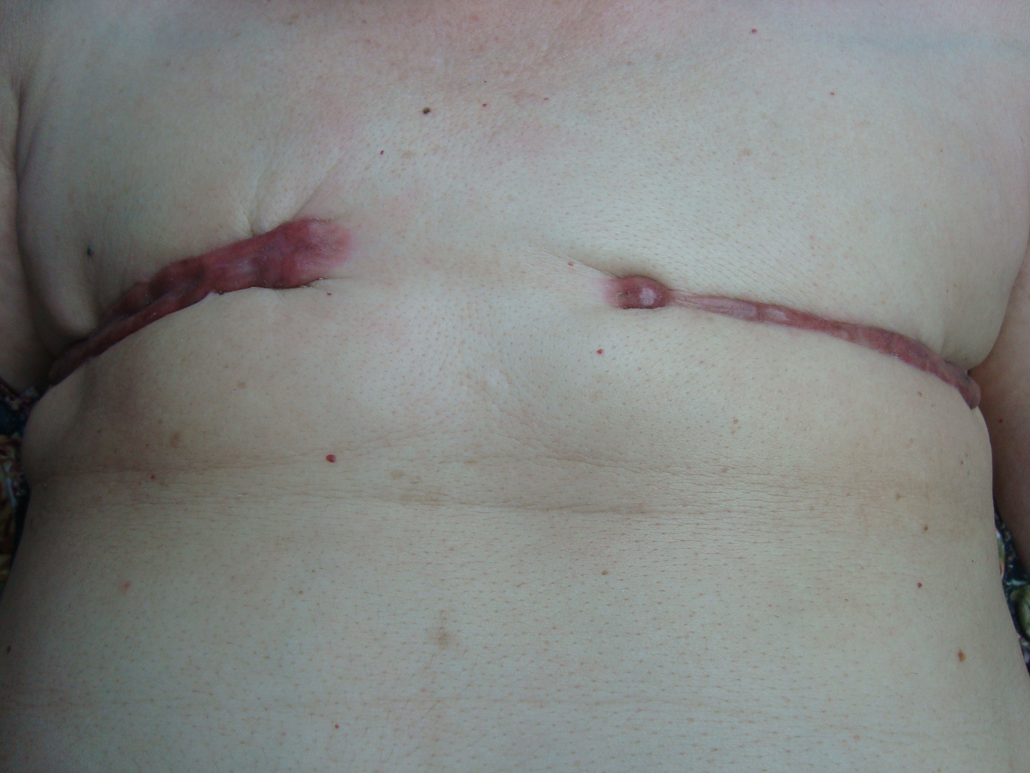
Another patient with bilateral modified radical mastectomies.  Note the hypertrophic scars to keloid formation.

DSC04505

Right mastectomy scar.

DSC04506

Left mastectomy scar.  Note the greater keloid formation near the sternum.

IMG_1327A1

Patient with left breast cancer with sternal keloid.

kyamko-1

Right after modified radical mastectomy using an oblique mastectomy incision.

mrm_scar_oblique_A1

Oblique mastectomy incision  scar.  Note the hypertrophic scar on the medial side.

DSC03362

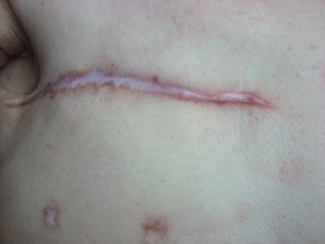
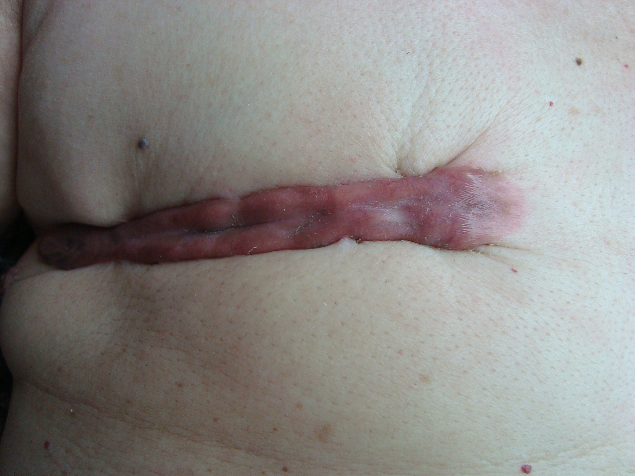
Transverse mastectomy incision. Note the hypertrophic scar on the medial side.

DSC03363

Closer-view of the transverse mastectomy incision scar with hypertrophic scar on the medial end.

DSC03508

A fine oblique mastectomy incision scar.

Posted in Keloid and Mastectomy | Leave a comment

2012 in review – ROJoson’s Blogs in WordPress.com

The WordPress.com stats helper monkeys prepared a 2012 annual report for this blog.

Here’s an excerpt:

The new Boeing 787 Dreamliner can carry about 250 passengers. This blog was viewed about 970 times in 2012. If it were a Dreamliner, it would take about 4 trips to carry that many people.

Click here to see the complete report.

Posted in Review of ROJoson's Blogs in Wordpress.com | Leave a comment

Difference between Fibrocystic Breast Changes and Fibroadenoma of the Breast on Operation and Palpation

Difference between Fibrocystic Breast Changes and Fibroadenoma of the Breast on Operation and Palpation

Difference between Fibrocystic Breast Changes and Fibroadenoma of the Breast on Operation and Palpation

Difference between Fibrocystic Breast Changes and Fibroadenoma of the Breast on Operation and Palpation

Fibroadenoma is a non-cancerous breast mass while fibrocystic breast changes are usually considered hormonal changes.

Picture above: Fibroadenoma – Note the presence of a whitish dominant mass with a clear border.

Picture above: Fibrocystic Breast Changes – Note the absence of a dominant mass; the whitish or pinkish breast tissues blend with the fatty tissues.

Correlating with the physical examination findings on palpation, fibrocystic changes usually present with a lumpy breast and fibroadenoma usually presents with a dominant mass.  Note: Depending on its characteristics, a dominant mass may be a fibroadenoma,  a macrocyst, phyllodes tumor, or even a cancer.

A palpable dominant mass with clear border which turns out to be a fibroadenoma.

The  appearance of the whole fibroadenoma right after removal, solid with well-defined and clear border.

The appearance of the fibroadenoma upon cutting it into half to look at the inner side which appears whitish and fibrous.

The appearance on operation of a fibrocystic breast change with no dominant mass, which is considered a normal breast tissue.

Other samples of fibrocystic breast changes – no dominant mass with blending of the whitish or pinkish areas with the yellow fatty tissues of the breast -again, to repeat, considered normal breast tissues.

In medical practice, fibrocystic breast changes are considered normal and do not need treatment, particularly operation.  However, sometimes, it may be hard to differentiate fibrocystic breast changes from breast cancers.  Thus, at times, patients are being operated on for dominant masses suspected to be cancers but which eventually turn out to be fibrocystic breast changes.

Posted in Fibrocystic Breast Changes vs Fibroadenoma | Leave a comment

Family history of breast cancer as a risk factor

Family history of breast cancer as a risk factor

Family history of breast cancer as a risk factor

Having a family history of breast cancer is a risk factor for a woman.

A risk factor is anything that increases a person’s chance of getting a disease, here, we are referring to breast cancer.

Having one first-degree relative (mother, sister, daughter) with breast cancer approximately doubles a woman’s risk, and having two first-degree relatives increases her risk fivefold.

Having  distant relatives (aunts, grandmothers, and cousins) with breast cancer slightly increases the risk.

Note that the majority of people diagnosed with breast cancer have NO family history of the disease.

+++++++++++++++++++++++++++++++++++++++++

Courtesy of:

http://sharing.mayoclinic.org/2012/10/10/breast-cancer-hits-home-three-times-in-one-year/#more-12066

Posted in Breast Cancer Consciousness, Breast Cancer Risk Assessment | Leave a comment

The likelihood of developing breast cancer within the next 10 years, by age

The likelihood of developing breast cancer within the next 10 years, by age

Posted on October 12, 2012

The likelihood of developing breast cancer within the next 10 years, by age

Posted on October 12, 2012

The likelihood of developing breast cancer within the next 10 years, by age:*

Age 20 0.6% 1 in 1,1760
Age 30 0.44% 1 in 229
Age 40 1.44% 1 in 69
Age 50 2.39% 1 in 42
Age 60 3.40% 1 in 29
Age 70 3.73% 1 in 27

*From American Cancer Society Breast Cancer Facts & Figures 2009-2010.

Posted in Breast Cancer Consciousness, Breast Cancer Risk Assessment | Leave a comment